
Pregnancy is full of quiet miracles and hidden risks. For most women, the occasional ultrasound scan offers reassuring glimpses of a growing baby. But for the roughly 10% of pregnancies complicated by issues such as fetal growth restriction, preeclampsia, or placental dysfunction, those brief snapshots can leave dangerous gaps. Blood flow between placenta and fetus can change rapidly; a single clinic visit may miss the difference between a temporary dip and a sustained problem that threatens oxygen and nutrient delivery. A new experimental device aims to close that gap.
Researchers, Engineers and clinicians from Stanford Medicine, the University of California San Diego (UC San Diego), and the University of Oxford jointly developed a wearable ultrasound patch, called “UPatch”, to continuously monitor fetal health.
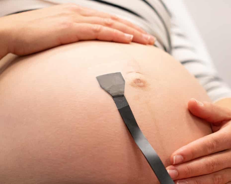
This flexible, adhesive wearable ultrasound patch nicknamed “UPatch” stays on the mother’s abdomen for hours and continuously tracks fetal anatomy and blood flow in real time, even as the baby and mother move. The work, published in Nature Biotechnology, represents a significant step toward continuous, autonomous prenatal monitoring that could make care more timely, precise, and personalized.
In Modern time, conventional obstetric ultrasound is powerful but limited. It requires a trained sonographer, specialized equipment, and a clinic or hospital setting. Even in high-risk cases, scans are typically performed only intermittently—sometimes once a day at most. Between appointments, clinicians rely on less specific tools such as cardiotocography which tracks fetal heart rate and contractions can produce false alarms or even miss subtle vascular problems.
The UPatch addresses these constraints by combining soft electronics, advanced ultrasound transducers, and intelligent software. Roughly the size of a palm, the soft, stretchable patch adheres to the abdomen. It uses arrays of miniature ultrasound transducers to generate and receive sound waves, producing both anatomical images (B-mode) and Doppler blood-flow spectra. Critically, the U-Patch uses real-time image-segmentation algorithms automatically to locate and follow target vessels such as the umbilical arteries and vein or the fetal middle cerebral artery without constant manual repositioning by a sonographer. This allows continuous acquisition of blood-flow waveforms even during fetal or maternal movement.
In validation studies, measurements from the patch closely matched those obtained with standard handheld clinical ultrasound devices across 62 pregnancies. Correlations were high for fetal heart rate and key Doppler indices such as the umbilical artery pulsatility index or cerebroplacental ratio, and biometric measurements (head circumference, abdominal circumference, femur length, estimated fetal weight) showed only small differences. Continuous sessions lasting one to six hours in 52 participants further demonstrated that the device could capture dynamic fluctuations in blood flow that a single snapshot would miss.
Seeing What Intermittent Scans Miss
One of the most compelling findings is the ability to distinguish transient changes from persistent compromise. Fetal blood-flow parameters can vary naturally over time. Continuous data revealed patterns that aligned with clinical diagnoses: healthy pregnancies showed expected gestational-age trends, while high-risk cases including preeclampsia, growth restriction, gestational diabetes and hypertension exhibiting distinctive Doppler signatures associated with placental insufficiency.
In a notable clinical example involving severe preeclampsia, the patch detected prolonged abnormal flow patterns consistent with significant intrauterine growth restriction. This information contributed to intensified monitoring and the decision for cesarean delivery at an earlier gestational age, potentially preventing stillbirth. Researchers emphasize that such continuous insight could allow earlier detection of conditions that currently rely on infrequent assessments.
The technology also measures multiple key vessels including all three major umbilical cord vessels (two arteries and one vein) plus important fetal arteries. Combined with anatomical imaging, it can support estimation of fetal size and growth trajectory which are critical metrics for diagnosing and managing growth restriction.
How the Engineering Works

Creating a wearable ultrasound device that works reliably on a moving fetus deep inside the body required solving several hard problems. Signal strength is weaker at greater depths; the umbilical cord and fetus constantly shift position; and the device must remain comfortable and safe for prolonged skin contact.
The team iterated through multiple prototypes, integrating high-performance piezoelectric transducers fabricated with specialized dicing techniques, acoustic lenses for focusing, and a soft Faraday cage for electromagnetic shielding. Soft, stretchable materials allow the patch to conform to the curved abdomen while maintaining acoustic coupling. Safety parameters stay within established limits set by regulatory and professional bodies for diagnostic ultrasound.
The software is equally important. Real-time segmentation and tracking algorithms keep the sample gate region from which Doppler data are extracted locked on the vessel of interest. This autonomy is what enables hours-long monitoring without a sonographer constantly adjusting the probe.
At present the system is still tethered by cable to external processing electronics which is a proof-of-concept stage. The researchers note that further miniaturization of the circuitry could enable fully wireless, more mobile versions.
If refined and validated in larger trials, continuous wearable ultrasound could shift high-risk pregnancy management from reactive to proactive.
Doctors might detect placental dysfunction or evolving fetal compromise earlier, tailor interventions more precisely, reduce unnecessary interventions driven by incomplete data, and potentially improve outcomes for both mother and baby. It could also expand monitoring access beyond specialized centers, especially if future wireless versions become practical for home or outpatient use under clinical supervision.
Nevertheless, the demonstration that a soft patch can autonomously track moving fetal vessels and deliver clinically meaningful continuous Doppler data is a genuine advance. It builds on broader progress in wearable ultrasound including earlier soft patches for other organs and brings the technology into one of medicine’s most sensitive domains.
Pregnancy monitoring has long been constrained by the trade-off between information richness and practicality. The UPatch suggests that trade-off can be improved. By turning occasional snapshots into a continuous stream, it offers the possibility of seeing fetal well-being not just in isolated moments, but across the ordinary, restless hours of pregnancy—when the most important changes may be happening.
Sources: Primary paper in Nature Biotechnology (Park et al., 2026) and contemporaneous reports from UC San Diego, Stanford Medicine, University of Oxford, and science outlets. All key claims in the original summary align with the published research.